Every chapter available in the Samacheer Kalvi Class 11th Bio Zoology Solutions subject is explained clearly in an easy way. Learn the depth concept by referring to the Samacheer Kalvi 11th Bio Zoology Solutions Chapter 9 Locomotion and Movement Questions and Answers. Have a look at every topic and get the complete knowledge on the Bio Zoology Solutions subject. You do not need to search for many materials for a better understanding of Bio Zoology Solutions. Just refer to Tamilnadu State Board Solutions pdf and have a grip on the total subject.
Tamilnadu Samacheer Kalvi 11th Bio Zoology Solutions Chapter 9 Locomotion and Movement
I believe that the best book is like a best friend to know the complete world by sitting in one place. When you have the best book you have many options to get great knowledge. Selecting the best book will lead to reaching your goal. Students who are looking for the best book to learn Bio Zoology Solutions can use Samacheer Kalvi Class 11th Bio Zoology Solutions Chapter 9 Locomotion and Movement Questions and Answers. Immediately start your learning with Samacheer Kalvi Class 11th Bio Zoology Solutions Solutions Pdf.
Samacheer Kalvi 11th Bio Zoology Locomotion and Movement Text Book Back Questions and Answers
Textbook Evaluation Solved
Question 1.
Muscles are derived from ………………..
(a) Ectoderm
(b) Mesoderm
(c) Endoderm
(d) Neuro ectoderm
Answer:
(b) Mesoderm
Question 2.
Muscles are formed by …………………
(a) Myocytes
(b) Leucocytes
(c) Osteocytes
(d) Lymphocytes
Answer:
(a) Myocytes
Question 3.
The muscles attached to the bones are called …………………
(a) Skeletal muscle
(b) Cardiac muscle
(c) Involuntary muscle
(d) Smooth muscles
Answer:
(a) Skeletal muscle
Question 4.
Skeletal muscles are attached to the bones by ………………….
(a) Tendon
(b) Ligament
(c) Pectin
(d) Fibrin
Answer:
(a) Tendon
Question 5.
The bundle of muscle fibres is called ………………….
(a) Myofibrils
(b) Fascicle
(c) Sarcomere
(d) Sarcoplasm
Answer:
(b) Fascicle
Question 6.
The pigment present in the muscle fibre to store oxygen is ……………………
(a) Myoglobin
(b) Troponin
(c) Myosin
(d) Actin
Answer:
(a) Myoglobin
Question 7.
The functional unit of a muscle fibre is …………………..
(a) Sarcomere
(b) Sarcoplasm
(c) Myosin
(d) Actin
Answer:
(a) Sarcomere
Question 8.
The protein present in the thick filament is …………………
(a) Myosin
(b) Actin
(c) Pectin
(d) Leucin
Answer:
(a) Myosin
Question 9.
The protein present in the thin filament is ……………….
(a) Myosin
(b) Actin
(c) Pectin
(d) Leucin
Answer:
(b) Actin
Question 10.
The region between two successive Z-discs is called a …………………
(a) Sarcomere
(b) Microtubule
(c) Myoglobin
(d) Actin
Answer:
(a) Sarcomere
Question 11.
Each skeletal muscle is covered by …………………
(a) Epimysium
(b) Perimysium
(c) Endomysium
(d) Hypomysium
Answer:
(a) Epimysium
Question 12.
Knee joint is an example of ………………..
(a) Saddle joint
(b) Hinge joint
(c) Pivot joint
(d) Gliding joint
Answer:
(b) Hinge joint
Question 13.
Name of the joint present between the atlas and axis is ……………….
(a) Synovial joint
(b) Pivot joint
(c) Saddle joint
(d) Hinge joint
Answer:
(b) Pivot joint
Question 14.
ATPase enzyme needed for muscle contraction is located in ………………
(a) Actinin
(b) Troponin
(c) Myosin
(d) Actin
Answer:
(c) Myosin
Question 15.
Synovial fluid is found in ……………..
(a) Ventricles of the brain
(b) Spinal cord
(c) Immovable joint
(d) Freely movable joints
Answer:
(d) Freely movable joints
Question 16.
Inflammation of joints due to accumulation of uric acid crystals is called as ……………..
(a) Gout
(b) Myasthenia gravis
(c) Dsteoporosis
(d) Osteomalacia
Answer:
(a) Gout
Question 17.
Acetabulum is located in ……………..
(a) Collar bone
(b) Hip bone
(c) Shoulder bone
(d) Thigh bone
Answer:
(b) Hip bone
Question 18.
Appendicular skeleton is ………………..
(a) Girdles and their limbs
(b) Vertebrae
(c) Skull and vertebral column
(d) Ribs and sternum
Answer:
(a) Girdles and their limbs
Question 19.
The type of movement exhibited by the macrophages are …………………
(a) Flagellar
(b) Ciliary
(c) Muscular
(d) Amoeboid
Answer:
(d) Amoeboid
Question 20.
The pointed portion of the elbow is ………………….
(a) Acromion process
(b) Glenoid cavity
(c) Olecranon process
(d) Symphysis
Answer:
(c) Olecranon process
Question 21.
Name the different types of movement?
Answer:
- Amoeboid movement
- Ciliary movement
- Flagellar movement
- Muscular movement
Question 22.
Name the filaments present in the sarcomere?
Answer:
Thick and thin filaments are the two types of filaments present inside the sarcomere.
Question 23.
Name the contractile proteins present in the skeletal muscle?
Answer:
Actin and myosin are contractile proteins present in the skeletal muscle.
Question 24.
When describing a skeletal muscle, what does “striated” mean?
Answer:
Each skeletal muscle fiber has a repeated series of dark and light bands. The dark A-bands and light I-bands give a striated appearance to the muscle.
Question 25.
How does an isotonic contraction take place?
Answer:
In isotonic contraction, the length of the muscle changes but the tension remains constant. The force produced is unchanged, e.g., lifting dumbbells and weight lifting.
Question 26.
How does an isometric contraction take place?
Answer:
In isotonic contraction, the length of the muscle changes but the tension remains constant. The force produced is unchanged, e.g., lifting dumbbells and weight lifting.
Question 27.
Name the bones of the skull?
Answer:
The skull is composed of two sets of bones – cranial and facial bones. It consists of 22 bones of which 8 are cranial bones and 14 are facial bones.
Question 28.
Which is the only jointless bone in the human body?
Answer:
The jointless bone is the hyoid bone in our throat. The hyoid bone (lingual bone) is a horseshoe.
Question 29.
List the three main parts of the axial skeleton?
Answer:
The skull, the vertebral column, and the ribcage are the three main parts of the axial skeleton.
Question 30.
How is tetany caused?
Answer:
Due to the deficiency of parathyroid hormone the level of calcium decreases in the blood that leads to rapid muscle spasm called tetany.
Question 31.
How does rigor mortis happen?
Answer:
After the death of an individual, the membrane of muscle cells becomes more permeable to calcium ions. This happens due to the partial contraction of skeletal muscles. The contracted muscles are unable to relax. This condition is known as rigor mortis.
Question 32.
What are the different types of rib bones that form the rib cage?
Answer:
- True rib bones (First 7 pairs)
- False rib bones (8, 9, 10th pairs)
- Floating rib bones (11 and 12th pair)
Question 33.
What are the bones that make the pelvic girdle?
Answer:
Ilium, ischium, and pubis make the pelvic girdle.
Question 34.
List the disorders of the muscular system?
Answer:
- Muscle fatigue
- Atrophy
- Muscle pull
- Muscular dystrophy
Arthritis
- Osteoarthritis
- Rheumatoid arthritis
- Gout
Question 35.
Explain the sliding-filament theory of muscle contraction?
Answer:
Andrew F.Huxley and Rolf Niedergerke proposed the sliding filament theory to explain muscle contraction. According to this theory, overlapping actin and myosin filaments of fixed length slide past one another in energy-requiring process, resulting in muscle contraction.
Question 36.
What are the benefits of regular exercise?
Answer:
- The benefits of regular exercise are:
- The muscles used in exercise grow larger and stronger.
- The resting heart rate goes down.
- More enzymes are synthesized in the muscle fiber.
- Ligaments and tendons become stronger.
- Joints become more flexible.
- Protection from a heart attack.
- Influences hormonal activity.
- Improves cognitive functions.
- Prevents obesity.
- Promotes confidence, esteem.
- Aesthetically better with a good physique.
- Overall well-being with good quality of life.
- Prevents depression, stress, and anxiety.
In-Text Questions Solved
Question 1.
Which myofilament has the binding sites for calcium? Name the specific molecule that binds with calcium?
Answer:
Actin filament has the binding sites for calcium. Troponin binds to calcium on thin filaments.
Question 2.
All muscles produce movement, but only skeletal muscle is responsible for locomotion. What is meant by this statement?
Answer:
All the muscles, skeletal, smooth, and cardiac, produce movement. Smooth muscles control the activities of internal organs like the intestine, stomach, lungs, bladder, etc. and their actions are involuntary. Cardiac muscles help in the functioning of the heart. But only skeletal muscles are responsible for locomotion.
Locomotion is effected by both bones and muscles attached to the bones and takes place by the coordination between muscular contraction and relaxation and the skeletal system. Moreover, the functioning of those skeletal muscles is voluntary.
Question 3.
The pelvic girdle is a heavy, strong girdle. How does its structure reflect its function?
Answer:
The pelvic girdle is a heavy structure specialized for weight-bearing. This feature enables it to connect the trunk and the legs, support and balance the trunk, and contain and supports the intestines, the urinary bladder, and the internal sex organs.
Question 4.
An exhausted student was attending a lecture. After 30 minutes or so, he lost interest and he let go with a tremendous yawn. To his great distress, he couldn’t close his mouth -his lower jaw was locked open. What do you think would have caused it?
Answer:
When he opened his mouth very wide, the mandibular condyle might have slid forward to the point that the joint might have got dislocated.
Samacheer Kalvi 11th Bio Zoology Locomotion and Movement Additional Questions & Answers
I. Choose The Correct Answer
Question 1.
The pseudopodia of amoeba are formed from
a) Cytoplasm
b) Nucleoplasm
c) Sarcoplasm
d) all the above
Answer:
a) Cytoplasm
Question 2.
The skeletal system is derived from
(a) Ectoderm
(b) Endoderm
(c) Mesoderm
(d) Mesoglea
Answer:
(c) Mesoderm
Question 3.
Name the movement of spermatozoa
a) amoeboid movement
b) flagellated movement
c) Ciliary movement
d) None of the above
Answer:
b) flagellated movement
Question 4.
The thick filament of muscle fibre is made up of ……………….
(a) Actin
(b) Myosin
(c) Tropomyosin
(d) Troponin
Answer:
(b) Myosin
Question 5.
The cytoplasm of the muscle fibre is called:
a) Myofibril
b) Sarcomere
c) Sarcoplasm
d) Sarcolemma
Answer:
c) Sarcoplasm
Question 6.
Where is the hyoid bone present?
(a) Cranium
(b) Appendicular skeleton
(c) Pectoral girdle
(d) Base of the buccal cavity
Answer:
(d) Base of the buccal cavity
Question 7.
Name the membrane which covers each fasciculus
a) Epimycium
b) Perirnycium
c) Endomyciurn
d) Mesomvscium
Answer:
b) Perirnycium
Question 8.
How many thoracic vertebrates are there?
(a) 7
(b) 12
(c) 5
(d) 4
Answer:
(b) 12
Question 9.
Rib cage protects ……………….
(a) Brain
(b) Kidney
(c) Lungs, heart, liver
(d) Heart
Answer:
(c) Lungs, heart, liver
Question 10.
The forearm bones are the ………………
(a) Tibia and fibula
(b) Radius and ulna
(c) Carpals and metacarpals
(d) Tarsal and metatarsals
Answer:
(b) Radius and ulna
Question 11.
Name the protein that regulates muscle contraction.
a) Tropomyosin and actin
b) Troponin and myosin
c) tropomyosin and troponin
d) None of the above
Answer:
c) tropomyosin and troponin
Question 12.
Which of the following allows movement in only one direction?
(a) Pivot joint
(b) Ball and socket joint
(c) Saddle joint
(d) Hinge joint
Answer:
(d) Hinge joint
Question 13.
Which chemical initiates the opening of multiple gated channels in sarcolemma?
a) Epinephrine
b) Norepinephrine
c) acetylcholine
d) erythromycin
Answer:
c) acetylcholine
Question 14.
Which of the following arthritis is related to protein metabolism?
(a) Osteoarthritis
(b) Rheumatoid arthritis
(c) Gouty arthritis
(d) Osteoporosis
Answer:
(c) Gouty arthritis
Question 15.
The skeletal system is formed from this layer!
a) Ectoderm
b) Mesoderm
c) Endoderm
d) Neuroderm
Answer:
b) Mesoderm
II. Fill in the blanks
Question 1.
The sperm cells show …………………… movement.
Answer:
Flagellar
Question 2.
Skeletal muscle is attached to the bone by a bundle of collagen fibres known as ……………………
Answer:
Tendon
Question 3.
The cytoplasm of the muscle fibre is called the ……………………
Answer:
Sarcoplasm
Question 4.
…………………… is a red coloured respiratory pigment of the muscle fibre.
Answer:
Myoglobin
Question 5.
…………………… are the granules of stored glycogen.
Answer:
Glycosomes
Question 6.
…………………… is the functional unit of the skeletal muscle.
Answer:
Sarcomere
Question 7.
The thick filaments are composed of the protein ……………………
Answer:
Myosin
Question 8.
The monomer of the myosin molecule is ……………………
Answer:
Meromyosin
Question 9.
The study of muscle is called ……………………
Answer:
Myology
Question 10.
The junction between the motor neuron and the sarcolemma of the muscle fibre is called the ……………………
Answer:
Neuromuscular junction or motor endplate
Question 11.
When a nerve impulse reaches a neuromuscular junction, …………………… is released.
Answer:
Acetylcholine
Question 12.
In …………………… contraction of the length of the muscle changes but the tension remains constant.
Answer:
Isotonic
Question 13.
In …………………… contraction of the length of the muscle does not change but the tension of the muscle changes.
Answer:
Isometric
Question 14.
The oxidative fibres are called …………………… fibres.
Answer:
Red muscle
Question 15.
Glycolytic fibres or white muscle fibres lack ……………………
Answer:
Myoglobin
Question 16.
The skeletal system is derived from the ……………………
Answer:
Mesoderm
Question 17.
The RBCs and WBCs are produced in the ……………………
Answer:
Bone marrow
Question 18.
The large hole in the temporal bone is the ……………………
Answer:
External auditory meatus
Question 19.
The lower jaw bone is called ……………………
Answer:
Mandible
Question 20.
…………………… is the only bone without any joint.
Answer:
Hyoid bone
Question 21.
The upper jaw is formed of the ……………………
Answer:
Maxilla
Question 22.
…………………… is the large opening found at the posterior base of the skull.
Answer:
Foramen magnum
Question 23.
Through foramen magnum, the medulla oblongata continues as the ……………………
Answer:
Spinal cord
Question 24.
The first vertebra is called the ……………………
Answer:
Atlas
Question 25.
The second vertebra is called the ……………………
Answer:
Axis
Question 26.
The cranium protects the ……………………
Answer:
Brain
Question 27.
…………………… is the flat bone on the midventral line of the thorax.
Answer:
Sternum
Question 28.
The first seven pairs of ribs are called ……………………
Answer:
True ribs/vertebro-sternal
Question 29.
The 11th and 12th pairs of ribs are called as …………………… ribs.
Answer:
Floating /vertebral
Question 30.
The 8th, 9th, and 10th pairs of ribs are called …………………… ribs.
Answer:
Flase /vertebro-chondral
Question 31.
Rib cage plays a role in ……………………
Answer:
Breathing
Question 32.
…………………… is a depression in the pectoral girdle on which the head of the humerus from the shoulder joint.
Answer:
Glenoid cavity
Question 33.
…………………… is situated at the upper end of the ulna which forms the pointed portion of the elbow.
Answer:
Olecranon process
Question 34.
…………………… is the largest, longest, and strongest bone in the body.
Answer:
Femur
Question 35.
The head of the femur articulates with the …………………… of the pelvis to form the hip joint.
Answer:
Acetabulum
Question 36.
The bone-forming cells are called ……………………
Answer:
Osteoblasts
Question 37.
The bone destroying cells are called the ……………………
Answer:
Osteoclasts
Question 38.
…………………… cells give rise to the osteoblasts.
Answer:
Osteogenic
Question 39.
The internal bone surfaces are covered with a delicate connective tissue membrane called the ……………………
Answer:
Endosteum
Question 40.
Between the epiphysis and diaphysis …………………… is present.
Answer:
Epiphyseal plate/growth plate
Question 41.
The …………………… are points of contact between the bones.
Answer:
Joints
Question 42.
Sutures of the flat skull bones are …………………… joints.
Answer:
Fibrous
Question 43.
…………………… are the freely movable joints.
Answer:
Synovial joints/Diarthroses joints
Question 44.
Between Atlas and axis …………………… joint is present.
Answer:
Pivot
Question 45.
Between the carpals …………………… joint is present.
Answer:
Plane/gliding
Question 46.
Between the carpal and metacarpal …………………… joint is present.
Answer:
Addle
Question 47.
Between humerus and pectoral girdle …………………… joint is seen.
Answer:
Ball and socket
Question 48.
…………………… is an autoimmune disorder affecting the action of acetylcholine.
Answer:
Myasthenia gravis
Question 49.
The removal of parathyroid gland results in ……………………
Answer:
Tetany
Question 50.
A traumatic pulling of the fibres produces a tear known as ……………………
Answer:
Sprain
Question 51.
…………………… is an inflammatory or degenerative disease that damages the joints.
Answer:
Arthritis
Question 52.
The wearing away of the bone ends of the knees and other movable joints are called ……………………
Answer:
Osteoarthritis
Question 53.
The inflammation of the synovial membranes is known as ……………………
Answer:
Rheumatoid arthritis
Question 54.
Inflammation of joints due to accumulation of uric acid crystals is known as ……………………
Answer:
Gout
Question 55.
The deficiency of vitamin D and hormonal imbalance cause ……………………
Answer:
Osteoporosis
Question 56.
The deficiency of vitamin D causes …………………… in children.
Answer:
Rickets
Question 57.
The deficiency of vitamin D causes …………………… in adults.
Answer:
Osteomalacia
Question 58.
…………………… increase the breathing and heart rate.
Answer:
Endurance or aerobic activities
Question 59.
…………………… protects us from a heart attack.
Answer:
Exercise
III. Short Answer Questions
Question 1.
What is amoeboid movement?
Answer:
The movement of cells by streaming movements of the cytoplasm forming pseudo-podia is known as amoeboid movement, e.g., macrophages.
Question 2.
What is ciliary movement?
Answer:
The movement caused by the cilia is ciliary movement, e.g., ciliated epithelial cells of respiratory passage and genital tracts.
Question 3.
Name the muscle protein?
Answer:
- Actin
- Myosin
Question 4.
What is muscular movement?
Answer:
The movement of hands, legs, jaws, tongue is caused by the contraction and relaxation of the muscle which is known as a muscular movement.
Question 5.
Classify the muscles on the basis of their rate of shortening?
Answer:
- Fast contraction fibre
- Slow contraction fibre
Question 6.
What are myofibrils?
Answer:
Each muscle fiber contains hundreds to thousands of rod-like structures called myofibrils that run parallel to its length.
Question 7.
How are muscles classified on the basis of ATP are activity?
Answer:
- Fast oxidative fibres
- Slow oxidative fibres
Question 8.
What is perimysium?
Answer:
The connective tissue covering around each fascicle is the perimysium.
Question 9.
What is sarcoplasm?
Answer:
The cytoplasm of the muscle fibre is called the sarcoplasm.
Question 10.
Distinguish between voluntary muscle and involuntary muscle.
Answer:
Voluntary muscle | Involuntary muscle |
| 1. The muscle whose functions are under the control of our will is a voluntary muscle. | 1. The muscle whose functions are not under the control of our will is the involuntary muscle. |
| 2. It is striated and hence called striated or striped muscle. | 2. It has no stripes and hence it is called smooth muscles or unstriped or non-striated muscles. |
Question 11.
What is meant by exoskeleton?
Answer:
It is a rigid hard case present outside the body of animals (eg) Cockroach.
Question 12.
What is myoglobin?
Answer:
Myoglobin is a red-coloured respiratory pigment of the muscle fibre.
Question 13.
What are the two types of endoskeletons?
Answer:
- Axial skeleton.
- Appendicular skeleton.
Question 14.
What are Glycosomes?
Answer:
Glycosomes are the granules of stored glycogen that provide glucose during the period of muscle fiber activity.
Question 15.
What is a brainbox?
Answer:
The cranial bones form the hard protective outer covering of the brain and called the brain box.
Question 16.
Distinguish between thick filaments and thin filaments?
Answer:
Thick filaments | Thin filaments |
| 1. The thick filaments are composed of the protein myosin. | 1. The thin filaments are composed of protein actin. |
Question 17.
Give notes on Jawbones?
Answer:
- It is composed of the upper jaw which is formed of the maxilla and the lower jaw is formed of the mandible.
- The upper jaw is fused with the cranium and is immovable.
- The lower jaw is connected to the cranium and is movable.
Question 18.
Name the proteins which regulate the contraction of muscles?
Answer:
Actin, myosin, tropomyosin, and troponin.
Question 19.
What is myology?
Answer:
The study of muscle is called myology.
Question 20.
What are oxidative fibres?
Answer:
The muscle fibres that contain numerous mitochondria and have a high capacity for oxidative phosphorylation are classified as oxidative fibres. They are also called red muscle fibres.
Question 21.
What are glycolytic fibres?
Answer:
The muscle fibres which contain a few mitochondria but possess a high concentration of glycolytic enzymes and large stores of glycogen are called glycolytic fibres. These lack myoglobin and hence these are pale. These muscle fibres are termed as white muscle fibres.
Question 22.
What is a hydrostatic skeleton?
Answer:
The skeleton found in soft-bodied invertebrates is called a hydrostatic skeleton. It is a fluid-filled cavity encircled by muscles, e.g., the earthworm.
Question 23.
Distinguish between exoskeleton and endoskeleton.
Answer:
Exoskeleton | Endoskeleton |
| 1. The rigid hard case present outside the body of animals is the exoskeleton. | 1. The rigid support structure found inside the body of vertebrates is the endoskeleton. |
| 2. e.g., Cockroach | 2. e.g., Human being |
Question 24.
What is a hydrostatic skeleton?
Answer:
The skeleton found in soft-bodied invertebrates is called a hydrostatic skeleton. It is a fluid-filled cavity encircled by muscles, e.g., the earthworm.
Question 25.
What is periosteum?
Answer:
The external surface of the bone is covered by a double-layered membrane called the periosteum. What is endosteum?
Question 26.
What are false ribs?
Answer:
The 8th, 9th, and 10th pairs of ribs do not articulate directly with the sternum but joined with the seventh rib.
Question 27.
What is the sternum?
Answer:
The sternum is a flat bone on the midventral line of the thorax. It provides space for the attachment of the thoracic ribs and abdominal muscles.
IV. Give Long Answers
Question 1.
Describe the different types of movements with examples.
Answer:
Types of Movements:
- Amoeboid movements
- Ciliary movements
- Flagellar movements
- Muscular movements
1. Amoeboid movements:
Cells such as macrophages exhibit amoeboid movement for engulfing pathogens by pseudopodia formed by the streaming movement of the cytoplasm.
2. Ciliary movements :
This type of movement occurs in the respiratory and genital passages.
3. Flagellar movement:
This type of movement occurs in the cells which are having flagella or whiplike motile organelles. (eg) Sperm cells.
4. Muscular movement:
The movement of the hand’s legs jaws is caused by the contraction and relaxation of the muscle.
Question 2.
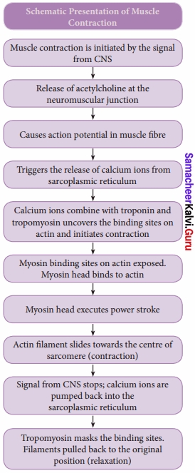
Write the schematic presentation of muscle contraction?
Answer:
Question 3.
Explain the structure of contractile proteins?
Answer:
The thick filaments of the muscle have the protein myosin. Each myosin molecule is made up of a monomer called meromyosin. It has a globular head, a short arm, and a tail. The short arm constitutes the heavy meromyosin (HMM). The tail portion forms the light meromyosin (LMM). The head bears an actin-binding site and an ATP- binding site. It also contains ATPase enzymes that split ATP to generate energy for the contraction of the muscle.
The thin filaments are composed of two intertwined actin molecules. It has polypeptide subunits called globular actin or G-actin and filamentous form or F-actin.
Each thin filament is made of two F-actins helically wound to each other. Each F-actin is a polymer of monomeric G-actins. Tropomyosin and troponin are other proteins which help in regulating the contraction of muscles along with actin and myosin.
Question 4.
Explain the types of skeletal muscle fibres?
Answer:
Skeletal muscle fibres are classified into three types. They are:
- Slow – oxidative fibres
- Fast – oxidative fibres
- Fast – glycolytic fibres
1. Slow-oxidative fibres have low rates of myosin ATP hydrolysis but have the ability to make large amounts of ATP. These fibres are used for prolonged, regular activity such as long-distance swimming. Long-distance runners have a high proportion of these fibres in their leg muscles.
2. Fast – oxidative fibres have high myosin ATPase activity and can make large amounts of ATP. They are particularly suited for rapid actions.
3. Fast – glycolytic fibres have myosin ATPase activity but cannot make as much ATP as oxidative fibres, because their source of ATP is glycolysis. These fibres are best suited for rapid, intense actions, such as short sprint at maximum speed.
Question 5.
Give the four important features of skeletal muscles.
Answer:
- Excitability: Based on the chemical and electrical excitation the muscles contract.
- Contractility: It is the ability of the muscle which gives movement to the attached organs.
- Conductivity: The excitation at one part of the muscle is connected to the other part of the muscle.
- Elasticity: The ability of the muscle to returns to its original position after the extension of the muscles.
Question 6.
Explain the bones that form the skull?
Answer:
The skull is composed of two sets of bones – cranial and facial bones. It consists of 22 bones of which 8 are cranial bones and 14 are facial, bones. The cranial bones form the hard protective outer covering of the brain and called the brain box. The capacity of the cranium is 1500 cm3.
These bones are joined by sutures which are immovable. They are paired parietal, paired temporal and individual bones such as the frontal, sphenoid, occipital and ethmoid. The large hole in the temporal bone is the external auditory meatus. In the facial bones maxilla, zygomatic, palatine, lacrimal, nasal are paired bones whereas mandible or lower jaw and vomer are unpaired bones. They form the front part of the skull.
A single U-shaped hyoid bone is present at the base of the buccal cavity. It is the only bone without any joint. Each middle ear contains three tiny bones- malleus, incus, and stapes collectively are called ear ossicles. The upper jaw is formed of the maxilla and the lower jaw is formed of the mandible.
The upper jaw is fused with the cranium and is immovable. The lower jaw is connected to the cranium by muscles and is movable. The most prominent openings in the skull are the orbits and the nasal cavity. The foramen magnum is a large opening found at the posterior base of the skull. Through this opening, the medulla oblongata of the brain descends down as the spinal cord.
Question 7.
Give an account of the vertebral column.
Answer:
- It consists of 33 serially arranged vertebrae which are interconnected by a cartilage known as the intervertebral disc.
- The vertebral column extends from the base of the skull to the pelvis and forms the main framework of the trunk.
It has five major regions, they are
- Cervical – 7
- Thoracic vertebrae -12
- Lumbar vertebrae – 5
- Sacrum – 5 sacral vertebrae found in the infant are fused to form one bone in the adult.
- Coccyx – 1 – 4 Coccygeal vertebrae found in the infant which are fused to form one bone in the adult.
Question 8.
Write a short note on Rib cage?
Answer:
There are 12 pairs of ribs. Each rib is a thin flat bone connected dorsally to the vertebral column and ventrally to the sternum. It has two articulation surfaces on its dorsal end, hence called bicephalic.
The first seven pairs of ribs are called ‘true ribs or vertebro-stemal ribs. Dorsally they are attached to the thoracic vertebrae and ventrally connected to the sternum with the help of hyaline cartilages.
The 8th, 9th and 10th pairs of ribs do not articulate directly with the sternum but joined with the cartilaginous (hyaline cartilage) part of the seventh rib. These are called ‘false ribs’ or vertebro-chondral ribs.
The last 11th and 12th pairs of ribs are not connected ventrally. Therefore, they are called as ‘floating ribs’ or vertebral ribs. Thoracic vertebrae, ribs and sternum together form the ribcage. Rib cage protects the lungs, heart, liver and also plays a role in breathing.
Question 9.
Write a note on Pectoral girdle?
Answer:
The upper limbs are attached to the pectoral girdles. These are very light and allow the upper limbs a degree of mobility not seen anywhere else in the body. The girdle is formed of two halves. Each half of the pectoral girdle consists of a clavicle or collar bone and a scapula.
The scapula is a large, thin, triangular bone situated in the dorsal surface of the ribcage between the second and seventh ribs. It has a slightly elevated ridge called the spine which projects as a flat, expanded process called the acromion. The clavicle articulates with this process.
Below the acromion is a depression called the glenoid cavity which articulates with the head of the humerus to form the shoulder joint. Each clavicle is a long slender bone with two curvatures which lies horizontally and connects axial skeleton with appendicular skeleton.
Question 10.
Write a note on the bones of the upper limb?
Answer:
The upper limb consists of 30 separate bones and is specialized for1 mobility. The skeleton of the arm, the region between the shoulder and elbow is the humerus. The head of humerus articulates with the glenoid cavity of the scapula and forms the shoulder joint. The distal end of humerus articulates with the two forearm bones the radius and ulna. The forearm is the region between the elbow and the wrist.
Olecranon process is situated at the upper end of the ulna which forms the pointed portion of the elbow. The hand consists of carpals, metacarpals and phalanges. Carpals, the wrist bones, 8 in number are arranged in two rows of four each.
The anterior surface of the wrist has the tunnel-like appearance, due to the arrangement of carpals with the ligaments. This tunnel is termed as carpal tunnel. Metacarpals, the pajm bones are 5 in number and phalanges the digits bones are 14 in number.
Question 11.
Explain the structure of Pelvic Girdle?
Answer:
The pelvic girdle is a heavy structure specialized for weight bearing. It is composed of two hip bones called coxal bones that secure the lower limbs to the axial skeleton. Together, with the sacrum and coccyx, the hip bones form the basin-like bony pelvis.
Each coxal bone consists of three fused bones, ilium, ischium and pubis. At the point of fusion of ilium, ischium, and pubis a deep hemispherical socket called the acetabulum is present on the lateral surface of the pelvis.
It receives the head of the femur or thigh bone at the hip joint and helps in the articulation of the femur. Ventrally the two halves of the pelvic girdle meet and form the pubic symphysis containing fibrous cartilage..
The ilium is the superior flaring portion of the hip bone. Each ilium forms a secure joint with the sacrum posteriorly. The ischium is a curved bar of bone.
The V-shaped pubic bones articulate anteriorly at the pubic symphysis. The pelvis of male is deep and narrow with larger heavier bones and the female is shallow, wide and flexible in nature, and this helps during pregnancy which is influenced by female hormones.
Question 12.
Write a note on the bones of lower limb?
Answer:
The lower limb consists of 30 bones which carries the entire weight of the erect body and is subjected to exceptional forces when we jump or run. The bones of the lower limbs are thicker and stronger than the upper limbs.
The three segments of each.lower limb are the thigh, the leg or the shank and the foot. The femur is the single bone of the thigh. It is the largest, longest and strongest bone in the body.
The head of femur articulates with the acetabulum of the pelvis to form the hip joint. Two parallel bones, the tibia and fibula, form the skeleton of the shank.
A thick, triangular patella forms the knee cap, which protects the knee joint anteriorly and improves the leverage of thigh muscles acting across the knee. The foot includes the bones of ankle, the tarsus, the metatarsus and the phalanges or toe bones.
The foot supports our body weight and acts as a lever to propel the body forward, while walking and running. The tarsus is made up of seven bones called tarsals. The metatarsus consists of five bones called metatarsals. The arrangement of the metatarsals is parallel to each other. There are 14 phalanges in the toes which are smaller than those of the fingers.
Question 13.
Explain the structure of a typical long bone?
Answer:
A typical long bone has a diaphysis, epiphyses and membranes. A tubular diaphysis or shaft, forms the long axis of the bone. It is constructed of a thick collar of compact bone that surrounds a central medullary cavity or marrow cavity. The epiphyses are the bone ends.
Compact bone forms the exterior of epiphyses and their interior contains spongy bone with red marrow. The region where the diaphysis and epiphyses meet is called the metaphysis. The external surface of the entire bone except the joint surface is covered by a double-layered membrane called the periosteum.
The outer fibrous layer is dense irregular connective tissue. The inner osteogenic layer consists of osteoblasts (bone- forming cells) which secrete bone matrix elements and osteoclasts (bone-destroying cells). In addition, there are primitive stem cells, osteogenic cells, that give rise to the osteoblasts.
The periosteum is richly supplied with nerve fibres, lymphatic vessels and blood vessels. Internal bone surfaces are covered with a delicate connective tissue membrane called the endosteum. The endosteum covers the trabeculae of spongy bone and lines the canals that pass through the compact bone. It also contains both osteoblasts and osteoclasts. Between the epiphysis and diaphysis epiphyseal plate or growth plate is present.
Question 14.
What are joints?
Answer:
The joints are points of contact between bones.
Question 15.
Explain the types of joints?
Answer:
(I) Fibrous joints or Synarthroses: They are immovable fixed joints in which no movement between the bones is possible. Sutures of the flat skull bones are fibrous joints.
(II) Cartilaginous joints or Amphiarthroses: They are slightly movable joints in which the joint surfaces are separated by a cartilage and slight movement is only possible, e.g., Joints of adjacent vertebrae of the vertebral column.
(III) Synovial joints or Diarthroses joints: They are freely movable joints, the articulating bones are separated by a cavity which is filled with synovial fluid e.g., Pivot joint – between atlas and axis plane/gliding joint – between the carpals
- Saddle j oint – between the carpal and metacarpal
- Ball and socket joint – between humerus and pectoral girdle
- Hinge joint – knee joint
- Condyloid or angular or ellipsoid-joint between radius and carpal.
Question 16.
Write a short note on myasthenia gravis?
Answer:
Myasthenia gravis: An autoimmune disorder affecting the action of acetylcholine at neuromuscular junction leadihg to fatigue, weakening and paralysis of skeletal muscles. Acetylcholine receptors on the sarcolemma are blocked by antibodies leading to weakness of muscles. When the disease progresses, it can make chewing, swallowing, talking and even breathing difficult.
Question 17.
Explain muscle fatigue?
Answer:
Muscle fatigue is the inability of a muscle to contract after repeated muscle contractions. This is due to lack of ATP and accumulation of lactic acid by anaerobic breakdown of glucose.
Question 18.
Explain muscle atrophy?
Answer:
A decline or cessation of muscular activity results in the condition called atrophy which results in the reduction in the size of the muscle and makes the muscle to become weak, which occurs with lack of usage as in chronic bedridden patients.
Question 19.
Write a short note on muscle pull?
Answer:
Muscle pull is actually a muscle tear. A traumatic pulling of the fibres produces a tear known as a sprain. This can occur due to the sudden stretching of muscle beyond the point of elasticity. Back pain is a common problem caused by muscle pull due to improper posture with static sitting for long hours.
Question 20.
Write a short note on muscular dystrophy?
Answer:
The group of diseases collectively called muscular dystrophy is associated with the progressive degeneration of skeletal muscle fibres, weakening the muscles and leading to death from lung or heart failure. The most common form of muscular dystrophy is called ‘ Duchene Muscular Dystrophy (DMD).
Question 21.
Write a short note on Rib cage?
Answer:
There are 12 pairs of ribs. Each rib is a thin flat bone connected dorsally to the vertebral column and ventrally to the sternum. It has two articulation surfaces on its dorsal end, hence called bicephalic.
The first seven pairs of ribs are called ‘true ribs or vertebro-stemal ribs. Dorsally they are attached to the thoracic vertebrae and ventrally connected to the sternum with the help of hyaline cartilages.
The 8th, 9th, and 10th pairs of ribs do not articulate directly with the sternum but joined with the cartilaginous (hyaline cartilage) part of the seventh rib. These are called ‘false ribs’ or vertebro-chondral ribs.
The last 11th and 12th pairs of ribs are not connected ventrally. Therefore, they are called ‘floating ribs’ or vertebral ribs. Thoracic vertebrae, ribs, and sternum together form the ribcage. The rib cage protects the lungs, heart, liver and also plays a role in breathing.
Question 22.
Explain the basic categories of exercise and physical activity?
Answer:
Exercise and physical activity fall into four basic categories. Endurance, strength, balance, and flexibility. Endurance or aerobic activities increase the breathing and heart rate. They keep the circulatory system healthy and improve overall fitness.
Strength exercises make the muscles stronger. They help to stay independent and cany out everyday activities such as climbing stairs and carrying bags.
Balance exercises help to prevent falls which is a common problem in older adults. Many strengthening exercises also improve balance.
Flexibility exercises help to stretch body muscles for more freedom of joint movements.
Hope all the information given regarding Class 11th Tamilnadu State Board Bio Zoology Solutions will help you to get good knowledge. For any queries, you can contact us and clear your doubts. Connect with us using the comment section. Also, we love your feedback and review. Get your Chapter Wise Samacheer Kalvi Class 11th Textbook Solutions for Bio Zoology Solutions Chapter 9 Locomotion and Movement Questions and Answers PDF start learning for the exam.